
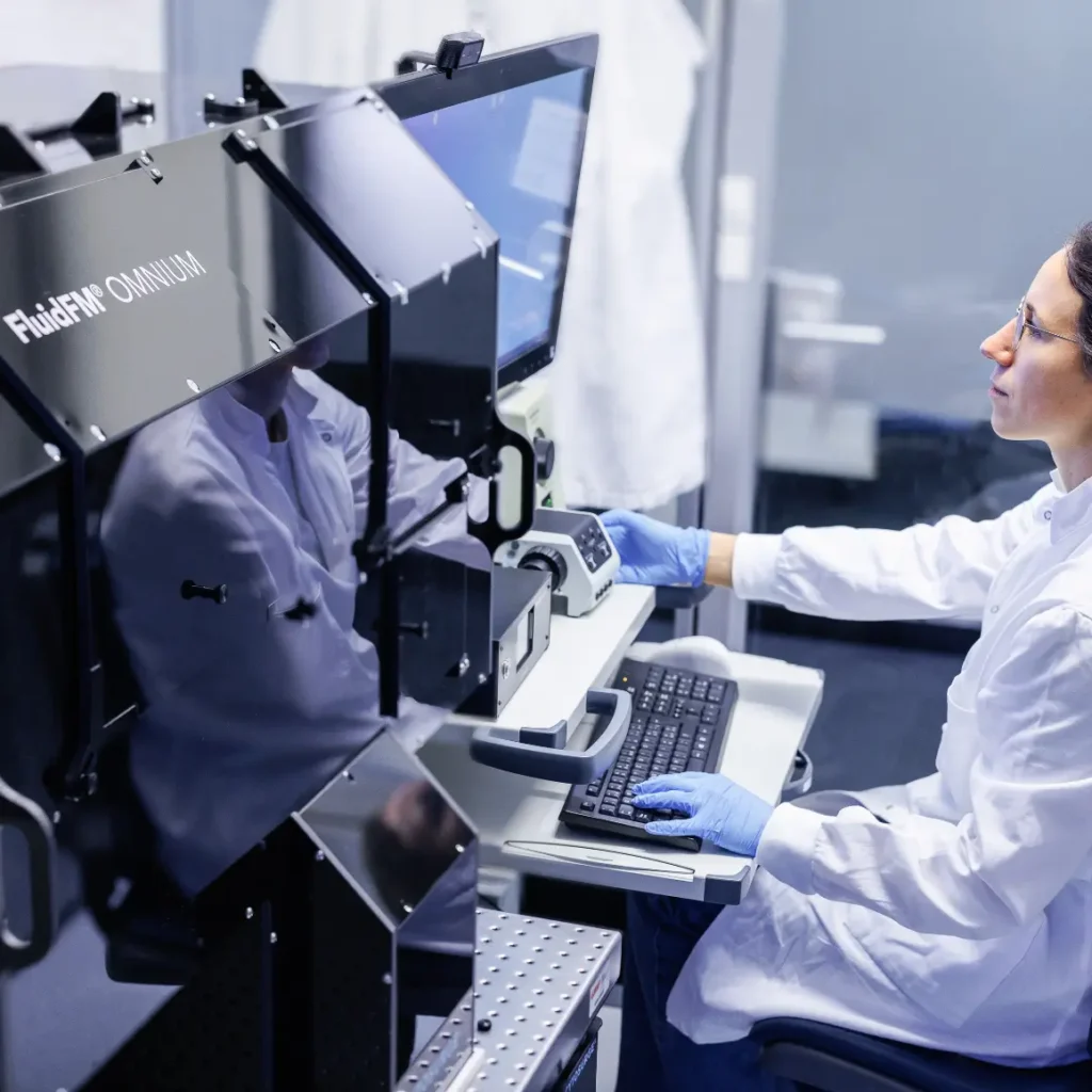

FluidFM® OMNIUM Platform

FluidFM® OMNIUM Platform

FluidFM® Technology – Combining Microfluidics and Force Microscopy

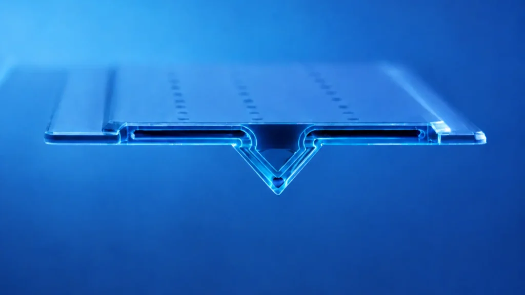
At the heart of the OMNIUM platform lies Cytosurge’s proprietary FluidFM technology, a unique approach that integrates closed microfluidic channels inside force-sensitive AFM probes. These specialized probes feature apertures as small as 300 nm, allowing them to aspirate or dispense femtoliter volumes of liquid while simultaneously measuring interaction forces down to the piconewton scale.

This dual capability provides exceptional control when interacting with biological samples. The integrated force feedback mechanism allows the system to precisely distinguish between gentle contact with the cell membrane and controlled membrane penetration, ensuring safe and reproducible experiments.

Thanks to this design, FluidFM enables a range of experimental operations that were previously extremely difficult or impossible with conventional methods.

Category:

Description

THE SAME CELL. BEFORE AND AFTER.

From Single-Cell Biology to Same-Cell Biology

Biology has traditionally relied on snapshots, measuring different cells at different moments and inferring causation from correlation. FluidFM® technology preserves cell viability during measurement, perturbation, or precise gene editing, revealing true cause and effect within the same cell.

This is Same-Cell Biology, a new approach to understanding how living cells behave, respond, and change.

FluidFM® Technology

FluidFM® technology combines a hollow AFM cantilever, a microfluidic channel, and force feedback into a probe for gentle manipulation of a single living cell without damaging it, with the precision to extract material or inject payloads, while the cell keeps living, dividing, and responding.

Specification

omplete OMNIUM full system specifications.

MetricSpecWhy it matters

Microscopy

Objectives

10x, 20x, 40x long-distance


Imaging

Brightfield


Configuration

Customizable


Focus stroke / increment

10.5 mm stroke, 0.01 µm increment

Fluorescence

Light source

White LED, 360–770 nm, 25,000 h lifetime


Filter sets

3 standard, Red, Green, Blue, expandable to 8


Filter switch

300 ms, motorized turret

Camera

Sensor

CMOS Color, 2.35 MP, 12-bit, 2nd camera option available

Incubation

Temperature

37°C ± 0.1°C


CO₂

1–15%, ± 0.1% accuracy


Sterility

HEPA filter + UV sterilization


Casing

Polycarbonate, optimized for fluorescence imaging

FluidFM

Pressure

−800 to +1,000 mbar, 0.5 mbar resolution


Stage

Nanometer-precise XY + Z, < 500 nm / < 5 nm repeatability


Probe exchange

Motorized, automated

Sample handling

Containers

Multi-well plates, up to 2x standard, dishes, glass-bottom dishes

Sample Compatibility: Your standard cell culture ware fits the system. No special consumables needed.

Expand Your System: Modular expansion with application-specific probes, consumables, and software. 

Applications

MULTIPLE SAME-CELL WORKFLOWS

One instrument for live-cell manipulation, measurement, and analysis

Use FluidFM OMNIUM across live-cell transcriptomics, vector-free delivery, cell isolation, force spectroscopy, and bioprinting workflows.

Live-seq

Repeated live-cell transcriptomics from the same living cell across time. Sample, perturb, and re-sample while preserving cell viability.

Learn more about Live-seq →

Vector-free cargo delivery

Deliver mRNA, proteins, CRISPR reagents, or nanomaterials directly into living cells without viral vectors or electroporation.

Learn more about CellEDIT →

Single-cell pick & place

Select, relocate, and organize individual living cells with image-verified precision for array generation, co-culture design, and population sorting.

Single-cell isolation

Isolate individual living cells for downstream analysis, monoclonal expansion, and image-verified single-cell workflows.

Automated force spectroscopy

Quantify cell adhesion, stiffness, and mechanical response at single-cell resolution with automated force measurements across large experimental datasets.

Bioprinting

Pattern biomolecules and living cells in defined spatial arrangements from sub-micron features to centimetre-scale constructs.